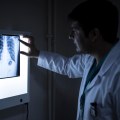
Radiology is a powerful tool for diagnosing a variety of diseases and conditions, such as Alzheimer's disease and dementia, anemia, appendicitis, arthritis and osteoporosis, blood clots and peripheral artery disease (PAD), brain tumors, many types of cancer, pneumonia, and chronic obstructive pulmonary disease (COPD). One of the most common types of scans is magnetic resonance imaging (MRI). An MRI can detect nerve injuries, tumors, brain injuries, strokes, or even the cause of a headache. Magnetic resonance imaging does not involve radiation, since it uses radio waves and magnetic fields to scan the body.
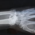
According to the National Institute for Biomedical Imaging and Bioengineering, X-rays are a form of ionizing radiation that can pass through most objects, including the human body. These advanced medical imaging techniques have many applications in diagnosing myocardial diseases, cancers of different tissues, neurological disorders, congenital heart diseases, abdominal diseases, complex bone fractures, and other serious medical conditions. In the future, with increasing innovations and advances in technological systems, the field of medical diagnostics will become a field for regular measurement of various complex diseases and will provide healthcare solutions. Many advanced techniques are being developed that can be explained by their operating principle, their application in medical laboratories and their development in diagnostic imaging techniques.
Medical imaging is the process of visually representing different tissues and organs in the human body to monitor the normal and abnormal anatomy and physiology of the body. Advanced medical imaging techniques include computed tomography (CT), positron emission tomography (PET), magnetic resonance imaging (MRI), single-photon emission computed tomography (SPECT), digital mammography and ultrasound. Contrast-enhanced dynamic magnetic resonance imaging (DCE-MRI) has been developed for the detection of the tumor microenvironment and its treatment. It is a new diagnostic tool that is used to detect diseases such as atherosclerosis, aging, cancer and schizophrenia, although it is still necessary to improve instrumentation and modeling for future purposes.
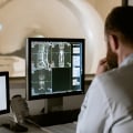
In the 1850s, many diagnostic tools such as ophthalmoscopes, stethoscopes and laryngoscopes made doctors think of the sensory power needed to develop other novel methods and techniques for diagnosing different diseases. However, medical ultrasound also had some side effects such as the effect of hormonal change, the breakdown of chromosomes at a very low frequency, chemical effects and other health problems. For the integration of big data into medical images researchers must develop algorithms to store images by converting them into a numerical format which will be useful for doctors in diagnosis. In the field of medicine technicians use computed tomography scanners machines to produce images that can diagnose anomalies and other therapeutic measures.
In the medical field ultrasound uses high-frequency sound waves to diagnose organs and body structure. With advanced medical imaging techniques it is possible to detect diseases at an early stage and ultimately help patients live better and longer lives. Medical imaging is the process of visually representing the structure and function of the different tissues and organs of the human body for clinical purposes and medical science for the detailed study of the normal and abnormal anatomy and physiology of the body.